Medical Imaging
At Coquille Valley Hospital, we believe that an investment in state-of-the-art technology is an investment in our community. Whether you are a patient or referring provider, we are here to answer your questions and provide the best possible imaging and service. Our goal is to offer high-quality, rapid testing and results that provide the answers needed to advance patient care.
Digital Mammography
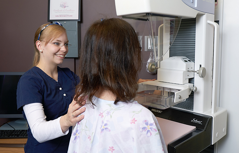
Compassionate and skilled, our medical imaging team ensures that our patients feel comfortable and are treated with the utmost respect and care. We offer the full range of breast imaging services, from mammograms to biopsies, to aid in the early detection and diagnosis of breast diseases. Mammography uses low-dose X-ray to examine the breasts and can be used for screening or diagnostic purposes. We offer state-of-the-art 2D and 3D technology using Hologic’s 3Dimensions mammography system, designed to provide high-quality images and a more comfortable experience for patients. For more information about mammography, regular screening and our latest imaging technology, visit our mammography page.
ACR Gold Seal of Accreditation for Mammography
Coquille Valley Hospital has been awarded a three-year term of accreditation in mammography by the American College of Radiology (ACR). The ACR gold seal of accreditation represents the highest level of image quality and patient safety. This is awarded only to facilities meeting ACR Practice Guidelines and Technical Standards after a peer-reviewed evaluation by board-certified physicians and medical physicists who are experts in the field. Image quality, personnel qualifications, adequacy of facility equipment, quality control procedures and quality assurance programs are assessed.
Preparing for a Mammogram
Little preparation is needed for a mammogram. Take your daily medications as you normally would, unless instructed otherwise. Wear two-piece clothing (top/bottom) and leave valuables at home, including jewelry. Avoid using deodorants, antiperspirants, powders, lotions, creams or perfumes under your arms or on your breasts. Metallic particles in powders and deodorants could be visible on your mammogram and cause confusion when reading the test results. You will be provided a gown to wear during your exam.
Bone Density Test – DEXA
A bone density test indicates if you have normal bone density, low bone density (osteopenia) or osteoporosis and is the only test that can diagnose osteoporosis. The lower your bone density, the greater your risk of breaking a bone. A bone density test, also called a DEXA scan, can help your healthcare provider develop a plan of care. Bone density tests are non-invasive and painless and usually take less than 15 minutes.
Preparing for a Bone Density Test
Patients are normally asked to avoid vitamin D and calcium 48 hours prior to the exam. With most types of bone density tests, the patient remains fully dressed. Clothing should be free of zippers, metal buttons, snaps or decorative jewelry. Please leave valuables at home, including jewelry.
CT
Also called computerized tomography, CT combines a series of X-ray views to produce cross-sectional images of the organs, bones and soft tissues inside your body. Once the information is collected, it can be reconstructed to view different angles. The resulting images can be compared to a loaf of sliced bread. Your doctor will be able to look at each of these “slices” individually or perform additional visualization to make 3D images. CT images provide much more information than standard X-rays and is especially helpful in emergency situations, such as a patient who may be suffering from internal injuries due to a car accident or other trauma. A CT scan can also visualize the brain and, with the help of injected contrast material, can check for blockages, blood vessel issues or other problems in the body.
Preparing for a CT Scan
Unless otherwise indicated, do not eat or drink anything but water four hours prior to your study. You may be asked to drink oral contrast before the test, which enhances the image quality. If your study requires contrast, we will indicate when to drink the contrast with specific instructions. Some studies require different preparations and do not require contrast. Please be sure to review the details for your particular study and contact us if you have questions. Please leave valuables at home, including jewelry.
MRI
Magnetic Resonance Imaging (MRI) uses a magnetic field and pulses of radio wave energy to make pictures of organs and structures inside the body. In many cases, MRI provides different information about structures in the body than can be seen with an X-ray, ultrasound or computed tomography (CT) scan. MRI also may show problems that cannot be seen with other imaging methods. For an MRI test, the area of the body being studied is placed inside a special machine that contains a strong magnet. In some cases, contrast material (gadolinium) may be used during the MRI scan to show certain structures more clearly.
Preparing for an MRI
There is little preparation needed for an MRI exam. Take your daily medications as you normally would, unless instructed otherwise. Wear loose-fitting clothing to your appointment and leave valuables at home, including jewelry. If your MRI requires contrast, you will be notified of additional requirements, such as withholding food and liquid intake. If you experience anxiety related to claustrophobia, your doctor may prescribe an oral medication for you to ease anxiety.
Ultrasound
Medical ultrasound, also known as diagnostic sonography or ultrasonography, is a diagnostic imaging technique based on the application of ultrasound. It is used to create an image of internal body structures, such as tendons, muscles, joints, blood vessels and internal organs. It is often used to find the source of a disease or to dismiss a suspected issue.
Preparing for an Ultrasound
Most ultrasound exams require no preparation; the exceptions are ultrasounds of the abdomen, pelvis, liver, aorta or kidneys, for which specific instructions will be provided. For some scans, such as a gallbladder ultrasound, your provider may ask that you not eat or drink for up to six hours before the exam, whereas a pelvic ultrasound may require a full bladder. You will be advised of any liquid or food intake requirements prior to your appointment. Please wear loose-fitting clothing and leave valuables at home, including jewelry.
X-ray
X-ray is a common imaging test that’s been used for decades. It can help your medical provider view the inside of your body to help diagnose, monitor and treat medical conditions. Different types of X-rays are used for different purposes. X-rays are commonly ordered to examine an area where a patient is experiencing pain or discomfort, a bone fracture, conditions affecting the lungs and heart and more.
Preparing for an X-ray
In most cases, you won’t need to take special steps to prepare for an X-ray, although, we suggest wearing loose, comfortable clothing. Please leave valuables at home, including jewelry. An X-ray technician will explain how and help you position your body and may ask you to lie, sit, or stand in a variety of positions during the test. It’s important to stay still while the images are being taken; this will provide the clearest images possible.
What Is The Patient Portal?
The patient portal is your online gateway to accessing medical information and communicating electronically. This is also a way to securely send and receive messages with providers and staff. View summaries of your visits, lab and imaging results, medications and more, and request and view upcoming appointments, all while keeping your information safe. You will need a code to access the online patient portal for the first time, which you can request at your first appointment.
Medical Imaging Hours
- Hours: Monday – Friday, 8:30 am – 5 pm
- Location: 940 E 5th St, Coquille, OR, 97423
- Directions: From Hwy 42, turn north onto N Central Blvd., turn east onto E 5th St; the hospital is on your left.
- Floor: 3rd
- Phone: 541-396-3101 Ext. 1057
- Fax: 541-396-6942